Common Conditions
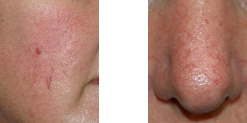
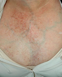
These veins and similar small vessels on the chest are usually due to sun damage. Facial veins may also be associated with flushing and vascular instability as seen in rosacea or carcinoid syndrome. Best treatment for these lesions is vascular laser therapy. Veins in the corner of the nose usually have higher flow volumes and are more difficult to treat. They are more common in people suffering with sinus disease and chronic nasal congestion. This can lead to recurrence despite adequate treatment. Vascular laser therapy may need to be repeated a number of times for these veins or injection sclerotherapy can be attempted.
Mat telangiectasias
These are patches of capillaries seen in patients with scleroderma and CREST syndrome. They present as flat and non-pulsatile vessels appearing as discrete mats. This should not be confused with telangiectatic matting which is a complication of surgery and sclerotherapy. Patients presenting with mat telangiectasias are screened for connective tissue disease and in particular scleroderma.
Hereditary Haemorrhagic Telangiectasias (HHT)

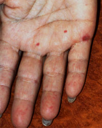
These lesions are arteriovenous malformations (AVM) of the microvasculature. They usually appear as small dark red elevated lesions on mucous membranes, face and distal limbs. Lesions may involve the tongue, retina, lung and the brain. HHT lesions appear during the third or fourth decade of life. Recurrent nose bleeds is usually the presenting symptom. Coagulation factor deficiency such as von Willebrand’s disease may be present. Family screening should be performed.
Ataxia Telangiectasia (AT)
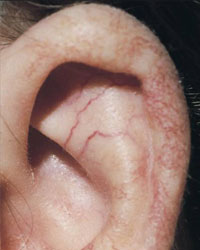
This is an autosomal recessive (inherited the bad gene from both parents) immunodeficiency (deficiency of the immune system). It is characterized by progressive neurologic impairment and cerebellar ataxia (loss of the ability to coordinate muscular movement). Patients are susceptible to lung and sinus infections and have a predisposition to certain cancers. Small capillaries are seen in the sclera of the eye and also on the helix of the ear. These veins appear later than the ataxia, first noticed in childhood and sometimes not until adolescence. The capillaries of the eye extend from the outside corner of the eye towards the centre. These small veins may also involve the ears, eyelids, inside of the elbows and behind the knees. The capillaries are predominantly of venous origin and are not AV malformations.
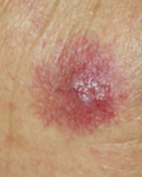
Post-Radiation
These small capillaries appear at the site of previos radiation treatment many years after radiotherapy. Skin cancers such as squamous cell carcinoma (SCC) can also develop in the same region.
Spider Naevi
These are dilated small arteries that arise from bigger and deeper arteries and travel straight up to the skin (ascending arterioles). They usually exhibit radiating thin-walled vessels in association with a central arteriole. These lesions are also referred to as spider angiomas and naevus araneus. Spider naevi are not growths but occur as a result of the expansion of preexisting vessels. They demonstrate arterial flow on Doppler examination. Compression of the central arteriole produces blanching. When released, the lesion quickly refills from the central arteriole. Spider naevi are not hereditary and are present in 10-15% of healthy adults and young children. Lesions are found most commonly on the face, neck, chest and arms. In young children, spider naevi are common on the backs of the hands and fingers.Hormonal changes play a role in the formation of these lesions. Many women develop lesions during pregnancy, while taking oral contraceptives, or after menopause. The lesions usually resolve spontaneously 6 to 9 months after delivery or after discontinuing oral contraceptives. Numerous lesions are seen in patients with significant liver diseasesuch as advanced hepatitis C.